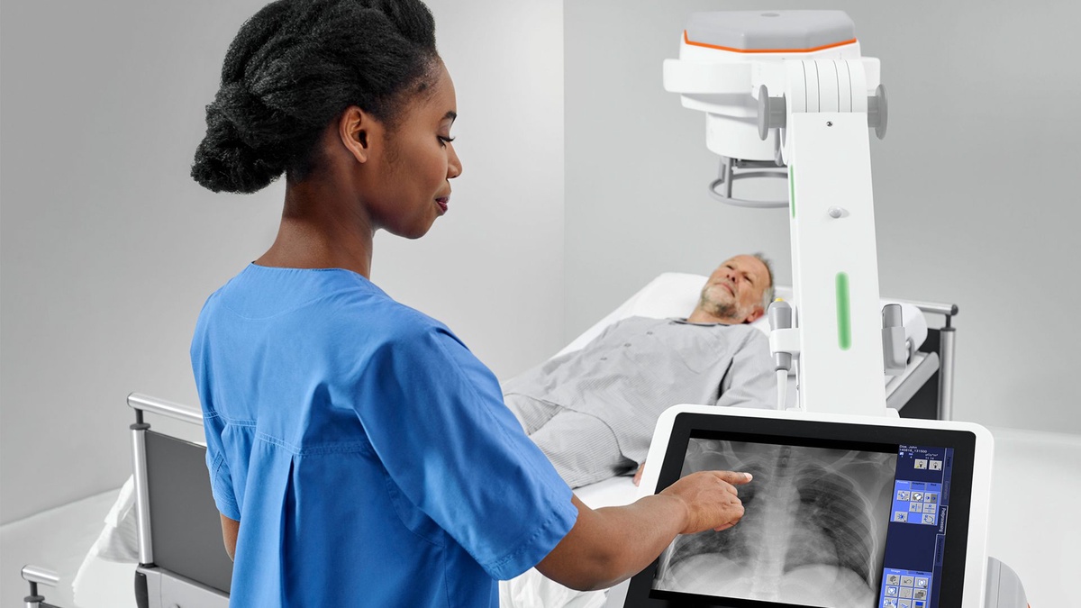
What is x-rays imaging?
Like visible light, X-ray imaging in Sacramento is electromagnetic radiation. In contrast to light, x-rays have more energy and can penetrate most materials, particularly the human body. To create images of the tissues and structures inside the body, medical x-rays are employed.
An image of the "shadows" generated by the things within the body will be created if x-rays passing through the body also pass through an x-ray detector on the patient's opposed side.
How do medical x-rays work?
An individual is positioned such that the body portion being scanned is situated between an x-ray source and an x-ray detector to generate a radiograph. When the equipment turns on, x-rays enter the body and, depending on the radiological density of the tissues they pass across, are absorbed in various amounts by various tissues.
The amount of radioactivity and atomic number (the quantity of protons in an atom's nucleus) of the substance being photographed are both factors that affect radiological density. For instance, calcium, found in human bones, has a greater atomic number than the majority of other organs.
This feature makes bones easily absorb x-rays, which results in strong contrast on the x-ray detector. As a result, against a radiograph's dark backdrop, bone structures stand out as being whiter than other tissues. In contrast, fat, muscle, and air-filled cavities like the lungs allow x-rays to pass through simpler because they are less radiologically dense than other types of tissue.
On a radiograph, these structures are seen in various hues of gray. When utilized properly, x-ray scans' diagnostic advantages vastly exceed their hazards. X-ray imaging Sacramento can identify illnesses that could be fatal, such as infections, cancer of the bone, and clogged blood veins. Ionizing radiation, a kind of radiation that x-rays create, has the ability to destroy living tissue.
Are there risks?
If the part of the body being photographed is not the abdomen or pelvis, there are no known dangers to the unborn child from an x-ray in a pregnant woman. In general, clinicians prefer to employ non-radiation tests like magnetic resonance imaging (MRI) or ultrasound when imaging the abdomen and pelvis is required.
However, an x-ray may be a suitable alternate imaging option if neither of them can offer the necessary answers, there is an emergency or another time limitation.
Conclusion
The x-ray tube is powered by electric current. The thermionic emission of electrons is made possible by the low voltage circuit attached to the cathode. The cathode to steel target electrons is accelerated by the high voltage circuit.
The majority of the kinetic energy of the electrons when they contact the target is converted to heat, and a very little amount is converted to an x-ray. Kilovoltage, which regulates the quality of the beam, milliamperage, and exposition time, which regulate the quantity of the beam, are the basic controls of the x-ray tube.
The radiation's intensity is determined by the separation between the x-ray source and the target. The radiographic image shows the various absorber densities, such as oral tissues.


No comments yet