Navigating the Maze of Brain Imaging:
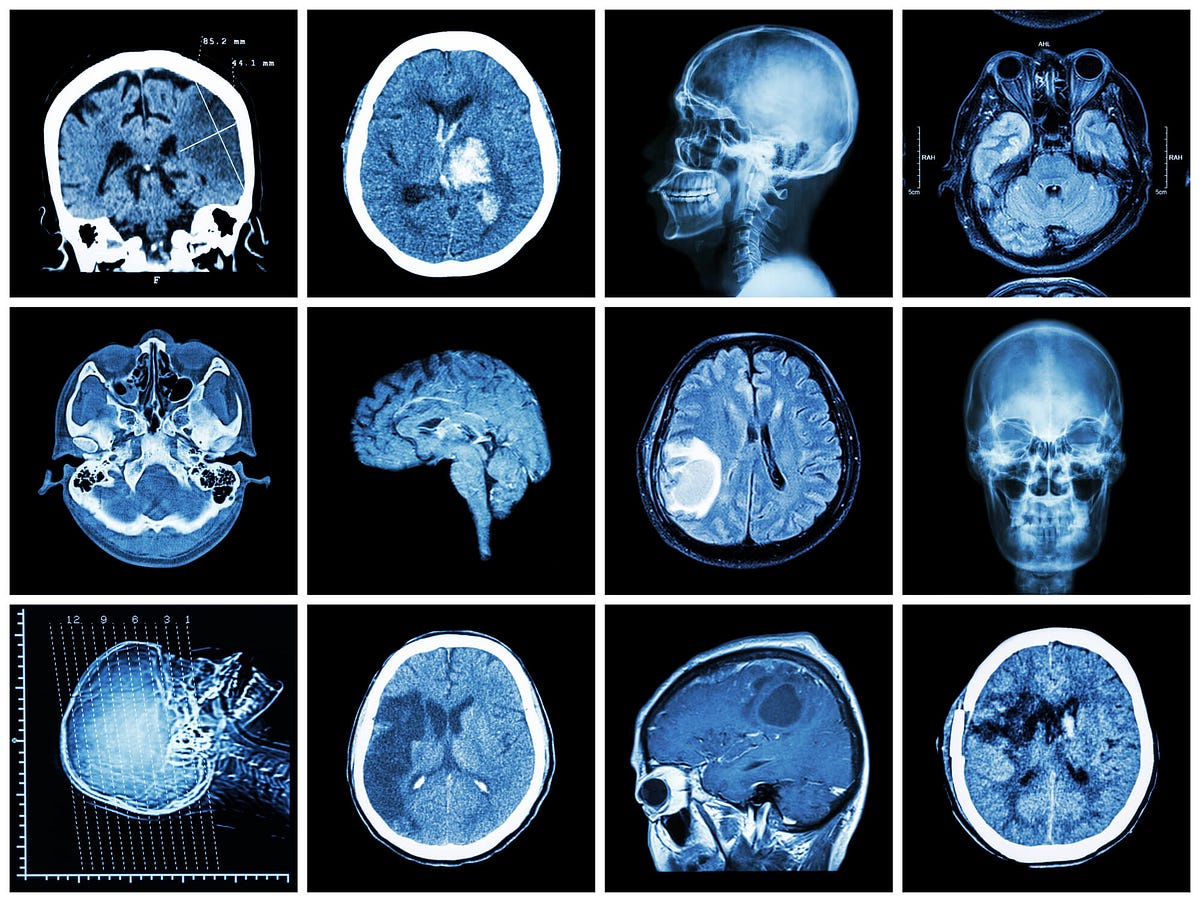
When it comes to assessing brain damage, modern medicine offers a variety of imaging techniques. From MRI and CT scans to PET scans, each method provides a unique perspective into the intricate workings of the brain. In this comprehensive guide, we aim to answer the pivotal question: Which scan shows brain damage?
Magnetic Resonance Imaging (MRI):
-
MRI, the master of details
If you're seeking a scan that captures the finer nuances of the brain's structure, an MRI is your go-to choice. This non-invasive imaging technique employs a powerful magnetic field and radio waves to generate detailed, high-resolution images of the Abnormality Brain scan soft tissues. Unlike CT scans, MRIs don't use ionizing radiation, making them a safer option for repeated use.
In the context of brain damage, MRIs excel at revealing abnormalities such as tumors, hemorrhages, and lesions. The detailed images produced by an MRI allow neurologists to identify the precise location, size, and nature of the damage, facilitating accurate diagnosis and treatment planning.
Computed Tomography (CT) Scan - Swift and Structural:
-
CT scans A quick overview
When time is of the essence, CT scans emerge as the frontrunners. These scans use X-rays to create cross-sectional images of the brain, providing a swift and efficient overview of its structure. CT scans are particularly valuable in emergency situations, such as detecting acute bleeding or skull fractures after a traumatic brain injury.
While CT scans excel in capturing structural abnormalities, they may not be as adept at revealing subtle changes in the brain's soft tissues. This makes them an excellent choice for emergencies but less suitable for comprehensive assessments of certain types of brain damage.
Positron Emission Tomography (PET) Scan - Functional Insights:
-
Exploring brain function with PET scans
If you're interested in gauging not just the structure but also the functional aspects of the brain, a PET scan is the way to go. PET scans involve injecting a small amount of a radioactive substance into the body, which is then detected by a special camera. This allows for the visualization of blood flow, oxygen consumption, and glucose metabolism in the brain.
In the realm of brain damage, PET scans are invaluable for assessing brain activity. They can reveal areas with reduced metabolic activity, indicating potential damage or dysfunction. This functional insight aids in understanding the impact of brain damage on cognitive processes, ultimately influencing treatment decisions.
When to Choose:
-
Weighing the pros and cons
The choice between MRI, CT, and PET scans often depends on the specific clinical scenario. In emergency situations, where quick insights into structural damage are crucial, a CT scan might be the initial choice. Its speed and efficiency make it indispensable in trauma cases.
However, for a more detailed and nuanced assessment, especially in non-emergency situations, an MRI takes the lead. Its ability to provide high-resolution images without radiation exposure makes it a preferred option for conditions requiring a closer look at the brain's soft tissues.
PET scans, on the other hand, shine in scenarios where understanding functional changes is paramount. This is particularly relevant in cases of neurodegenerative diseases or when evaluating the impact of brain damage on cognitive function.
Advancements on the Horizon:
-
The evolving landscape of brain imaging
As technology advances, so does the field of brain imaging. Emerging techniques such as functional MRI (fMRI) and diffusion tensor imaging (DTI) are pushing the boundaries of our understanding of Abnormality Brain Scan structure and function. These innovations hold the promise of even greater precision in diagnosing and monitoring brain damage.
Conclusion:
In the quest to understand which scan shows brain damage, the answer lies in recognizing the strengths and limitations of each imaging modality. MRI, with its detailed portrayal of soft tissues, CT scans, offering a swift structural overview, and PET scans, providing functional insights, collectively contribute to a holistic understanding of brain damage.
Medical professionals navigate this maze of imaging options based on the specific requirements of each case. The future holds exciting possibilities as technology continues to evolve, promising even more precise and personalized approaches to diagnosing and treating brain damage. Until then, the collaborative use of these imaging techniques remains essential in unraveling the complexities of the human brain.

No comments yet